MRI Imaging
Magnetic Resonance Imaging (MRI) is a non-invasive scanning method that produces detailed pictures of organs, bones, muscles, and blood vessels. Instead of ionizing radiation, it relies on a strong magnetic field paired with radio waves, which makes it a common alternative to CT scans when doctors need a closer look at soft tissue.
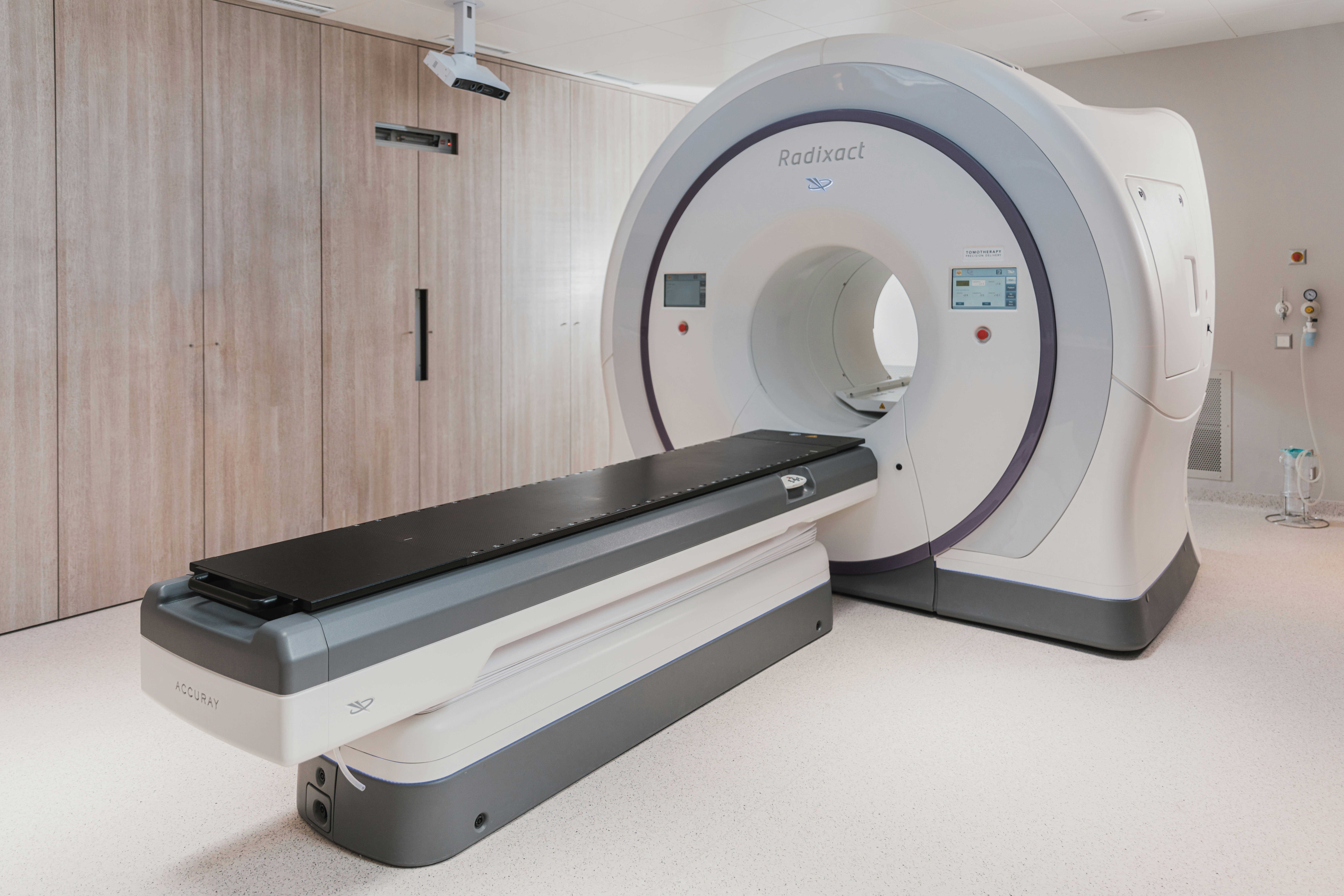
Medical radiation therapy machine in clinic. (2022, October). Unsplash. https://unsplash.com/photos/medical-radiation-therapy-machine-in-clinic-QTP8UKW_PgI
How it works
The scanner surrounds the body with a powerful magnetic field that briefly lines up the atoms in your tissue. Pulses of radio waves knock those atoms out of alignment, and as the pulses stop, the atoms settle back into place and give off a faint signal. A computer picks up that signal and reconstructs it into a cross-sectional image on a monitor.
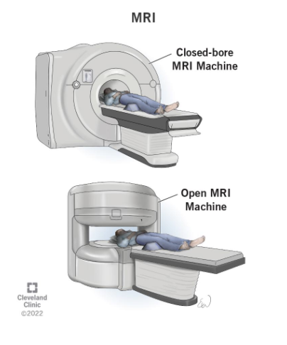
Open vs. closed scanners
Traditional closed MRI machines fully enclose the patient in a tube. They generally produce sharper images and finish faster, since their stronger magnets pick up more detail. The trade-off is comfort: the enclosed space can be difficult for people with claustrophobia or for larger patients.
Open MRI machines leave the sides uncovered, using magnets positioned above and below the patient instead of around them. This layout is easier on anxious or larger patients, but the weaker magnetic field usually means longer scan times and somewhat lower image resolution.
Other uses
- MRA (Magnetic Resonance Angiography): checks blood flow through arteries and can help spot aneurysms or vascular abnormalities.
- fMRI (Functional MRI): maps brain activity by having the patient perform a task, helping doctors locate the areas responsible for functions like speech or memory before surgery.
Getting ready for a scan
- Most patients can eat, drink, and take regular medication beforehand, though some specialty exams have their own restrictions.
- You'll be asked to remove clothing, jewelry, and anything metallic, and change into a gown.
- The scan takes place inside a tube-shaped machine open at both ends. Staying completely still is important for clear images, and earplugs are provided for the loud noise.
- If contrast dye is required and you've reacted to it before, let your doctor know in advance so you can be prescribed the right premedication.
- Anyone with anxiety about enclosed spaces can ask a doctor about anti-anxiety medication ahead of time — arrange for a ride home if you take it.
Important to disclose beforehand
Tell your doctor in advance if any of the following apply:
- A pacemaker or replaced heart valve
- An implanted pump, such as an insulin pump
- Vessel coils, filters, stents, or clips
- Pregnancy or possible pregnancy
- A past bullet wound
- A history of working with metal (e.g. grinding or welding)
- Metal fragments anywhere in the body
- Difficulty lying still for 30–60 minutes
Procedures for an MRI Scan
MRI scans may be performed on an outpatient (getting treatment without staying in an office overnight) basis or as part of a stay in a hospital. Although specific protocols may differ among facilities, an MRI procedure generally follows this process:
- You will be asked to remove all clothing, jewelry, eyeglasses, hearing aids, hairpins, removable dental work or other objects that may interfere with the procedure.
- You will be given a gown to wear.
- If you are to have a procedure done with contrast, an IV line will be started in the hand or arm for injection of the contrast dye. If the contrast is to be taken by mouth, you will be given the contrast to swallow.
- You will lie on a scan table that slides into a large circular opening of the scanning machine. Pillows and straps may be used to prevent movement during the procedure.
- The technologist will be in another room where the scanner controls are located. However, you will be in constant sight of the technologist through a window. Speakers inside the scanner will enable the technologist to communicate with and hear you. You will have a communication ball so that you can let the technologist know if you have any problems during the procedure. The technologist will be watching you at all times and will be in constant communication.
- You will be given earplugs or a headset to help block out the noise from the scanner. Some headsets may play music.
- During the scanning process, a clicking noise will sound as the magnetic field is created and pulses of radio waves are sent from the scanner.
- It will be important for you to remain very still during the examination, as any movement could cause distortion and affect the quality of the scan.
- At intervals, you may be instructed to hold your breath or not breathe for a few seconds, depending on the body part being examined. You will then be told when you can breathe. You should not need to hold your breath for longer than a few seconds.
- If contrast dye is used, you may experience some effects when it is injected into the IV line. These effects include a flushing sensation or a feeling of coldness, a salty or metallic taste in the mouth, a brief headache, itching, or nausea and/or vomiting. These effects usually last for a few moments.
- You should notify the technologist if you have any breathing difficulties, sweating, numbness or heart palpitations.
- Once the scan is complete, the table will slide out of the scanner and you will be helped off the table.
- If an IV line was inserted for contrast administration, the line will be removed.
- While the MRI procedure itself causes no pain, having to lie still for the length of the procedure might cause some discomfort or pain, particularly if you had a recent injury or invasive procedure, such as surgery. The technologist will use all possible comfort measures and complete the procedure as quickly as possible.
After the scan
Stand up slowly to avoid lightheadedness. If you received a sedative, plan to rest until it wears off and arrange another way home. Watch the IV site for redness or swelling, and contact your doctor promptly if you notice any signs of a reaction to contrast dye, such as itching, rash, or trouble breathing — treat difficulty breathing as an emergency. Otherwise, most people can return to their normal routine right away.
Why MRI machines are expensive to maintain
| Component | Why it wears out | Typical replacement interval |
|---|---|---|
| RF coils | Physical wear, cable damage, connector failures, accidental drops | Every 3–10 years, sooner with heavy use |
| Magnet | Requires ongoing cooling to maintain zero electrical resistance | — |
| Gradient coils | Electrical component aging and thermal stress | — |
| Liquid-helium cooling system | Pumps, filters, valves, and hoses wear down over time | — |