CT Overview
A CT (computed tomography) scan is a diagnostic imaging procedure that combines X-rays with computer technology to produce detailed images of the inside of the body — bones, muscles, fat, organs, and blood vessels alike. It goes well beyond what a standard X-ray can show.
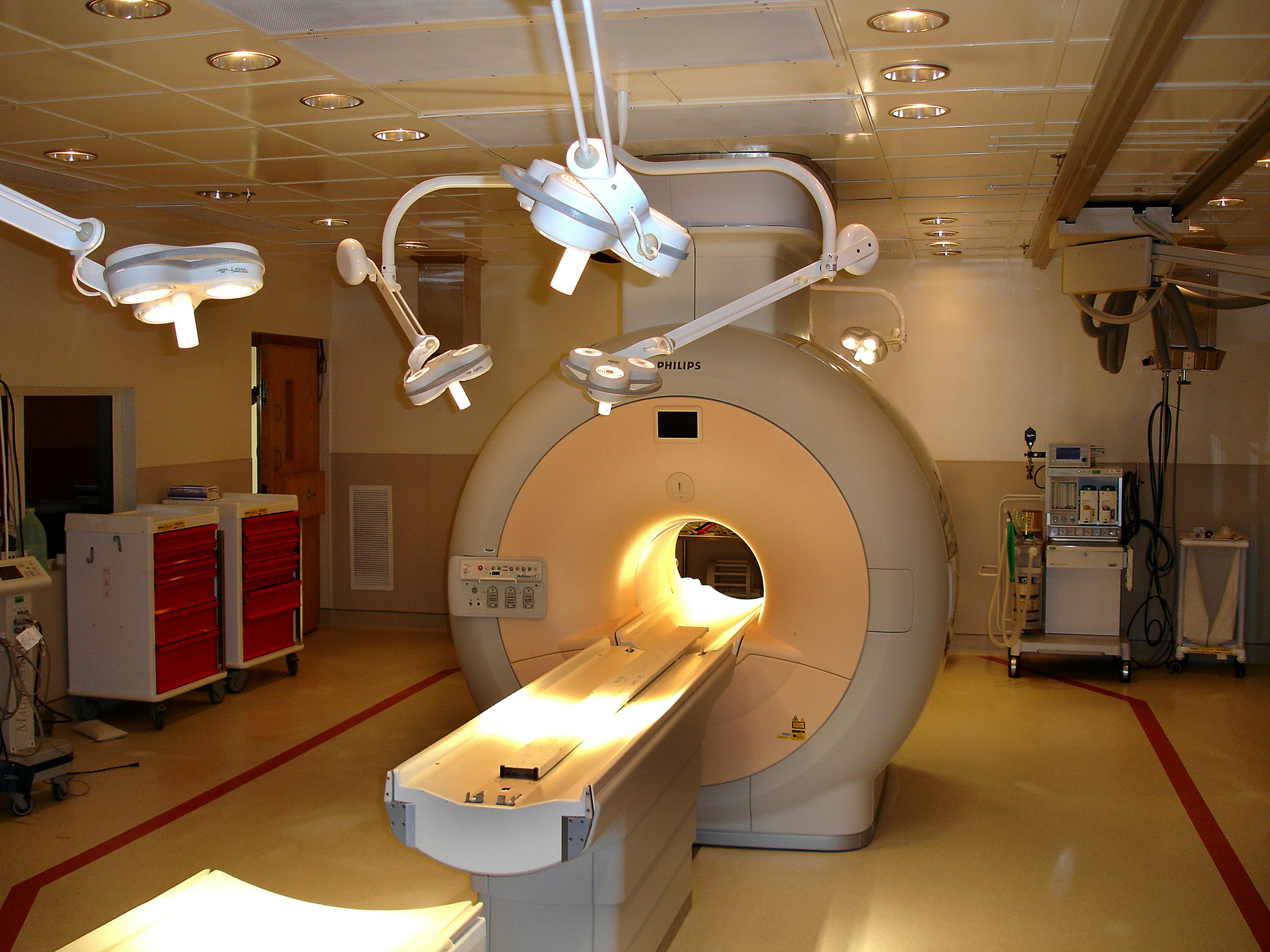
Trinks, D. (2023, August). A light that is inside of a room. Unsplash. https://unsplash.com/photos/a-light-that-is-inside-of-a-room-U9IriDpTcg4
How CT differs from a standard X-ray
In a standard X-ray, a single beam of energy passes through the body and a plate on the other side captures the variations left after it travels through skin, bone, muscle, and other tissue. That's useful, but it leaves a lot of internal detail unseen. In a CT scan, the X-ray beam instead rotates in a circle around the body, capturing many different views of the same structure. A computer interprets that data and assembles it into a two-dimensional image on a monitor; modern software can also reconstruct the data into three-dimensional images. CT scans are commonly used to help diagnose tumors, investigate internal bleeding, check for internal injuries, or guide a tissue or fluid biopsy.
Contrast
CT scans may be performed with or without contrast — a substance taken orally or injected through an IV that makes a particular organ or tissue easier to see clearly. Contrast exams sometimes require fasting beforehand. Patients should let their doctor know about any past reaction to contrast media or any kidney problems; a reported seafood allergy on its own is not considered a contraindication for iodinated contrast.
Preparing for a CT scan
- Pregnancy — patients who are or may be pregnant should check with their doctor before scheduling the exam.
- Clothing — a patient gown may be provided; piercings, jewelry, and valuables should be left at home.
- Contrast media — improves a radiologist's ability to spot abnormal structures and understand normal anatomy. Patients with kidney function issues should inform their radiologist in advance, since the scan may need to be done without contrast or via an alternate imaging method. A common approach — a double-contrast study — involves drinking a contrast media before the exam in addition to IV contrast; the more that's tolerated, the clearer the digestive tract appears on the images.
- Allergies — any past allergic reaction to contrast media should be reported when scheduling. IV contrast won't be given after a severe or anaphylactic reaction; milder past reactions are typically managed with pre-medication.
- Eating and drinking — no restrictions for scans without contrast. For scans with contrast, patients are usually asked not to eat for three hours beforehand, though clear liquids and prescribed medications are fine.
- Diabetes — a light meal three hours before the scan is typically recommended, and some oral diabetes medications may need to be paused for 48 hours afterward, per detailed instructions given at the time.
Risks
The radiation dose used in a CT scan is small, though patients can ask their doctor about the specific dose and risk for their situation. Patients who are pregnant or think they may be pregnant should notify their provider. Anyone who is claustrophobic or prone to anxiety should mention it ahead of time — a mild sedative can be prescribed to help.
Procedures for an CT Scan
CT scans may be performed on an outpatient basis or as part of your stay in a hospital. Procedures may vary depending on your condition and your physician's practices. Generally, CT scans follow this process, and the entire scan itself only takes a few minutes:
- You may be asked to change into a patient gown. If so, a gown will be provided for you. A locker will be provided to secure all personal belongings. Remove all piercings and leave all jewelry and valuables at home.
- If you are to have a procedure done with contrast, an IV line will be started in the hand or arm for injection of the contrast media, and the scan can begin immediately. For oral contrast, you will be given a liquid contrast preparation to swallow, which can take up to an hour to work through your system. In some situations, the contrast may be given rectally.
- You will lie on a scan table that slides into a large, circular opening of the scanning machine.
- The technologist will be in another room where the scanner controls are located. However, you will be in constant sight of the technologist through a window. Speakers inside the scanner will enable the technologist to communicate with and hear you. You may have a call button so that you can let the technologist know if you have any problems during the procedure. The technologist will be watching you at all times and will be in constant communication.
- As the scanner begins to rotate around you, X-rays will pass through the body for short amounts of time. You will hear clicking sounds, which are normal.
- The X-rays absorbed by the body's tissues will be detected by the scanner and transmitted to the computer. The computer will transform the information into an image to be interpreted by the radiologist.
- It is important that you remain very still during the procedure. You may be asked to hold your breath at various times during the procedure.
- If contrast media is used for your procedure, you may feel some effects when the contrast is injected into the IV line. These effects include a flushing sensation, a salty or metallic taste in the mouth, a brief headache, or nausea and/or vomiting. These effects usually last for a few moments.
- You should notify the technologist if you have any breathing difficulties, sweating, numbness or heart palpitations.
- When the procedure has been completed, you will be removed from the scanner.
- If an IV line was inserted for contrast administration, the line will be removed.
- While the CT procedure itself causes no pain, having to lie still for the length of the procedure might cause some discomfort or pain, particularly in the case of a recent injury or invasive procedure, such as surgery. The technologist will use all possible comfort measures and complete the procedure as quickly as possible to minimize any discomfort or pain.
After the scan
If contrast was used, patients may be monitored briefly for reactions like itching, swelling, rash, or difficulty breathing. Pain, redness, or swelling at the IV site after returning home should be reported to a doctor, since it could indicate infection. Generally, no special aftercare is needed — most patients can resume their usual diet and activities unless told otherwise.
Advances in CT technology
| Technology | What it does |
|---|---|
| High-resolution CT | Uses very thin slices (under 0.1 inches) for greater detail in conditions such as lung disease |
| Helical (spiral) CT | Patient and X-ray beam move continuously together, producing faster scans with greater resolution and contrast; multidetector versions can assess calcium buildup in the coronary arteries |
| Ultrafast (electron beam) CT | Captures images rapidly enough to create a "movie" of moving structures, such as the heart's chambers and valves; less common today than helical scanners |
| CT angiography (CTA) | Uses CT rather than standard X-ray or fluoroscopy to image blood vessels, such as the coronary arteries |
| PET/CT | Combines CT's detailed anatomy with PET's view of cell function and metabolism, aiding diagnosis of cancer, epilepsy, Alzheimer's disease, and coronary artery disease |
Why CT excels at imaging bone
- High contrast with surrounding tissue — bone's calcium content is dense and absorbs X-rays much more strongly than soft tissue, making fractures and bone abnormalities stand out clearly.
- Detailed 3D reconstruction — hundreds of X-ray images taken from different angles are combined into thin cross-sectional slices, which can be reconstructed into a 3D model for examining complex anatomy from any angle.
- Clarity on complex fractures — CT outperforms standard X-ray for fractures of the skull, spine, pelvis, face, and joints (ankle, wrist, knee, shoulder), and can reveal tiny fracture fragments and their exact alignment for surgical planning.
- Speed — most CT scans take only a few minutes, making CT the preferred imaging method for trauma patients with possible multiple injuries.