PET Overview
A positron emission tomography (PET) scan is an imaging test that shows organs and tissues at work. It uses a safe, injectable radioactive substance called a radiotracer along with a PET scanner, which detects cells absorbing large amounts of the tracer — a sign of potential disease. Because it's tracking activity rather than just structure, PET can measure vital functions like blood flow, oxygen use, and metabolism, and can catch disease at very early stages.
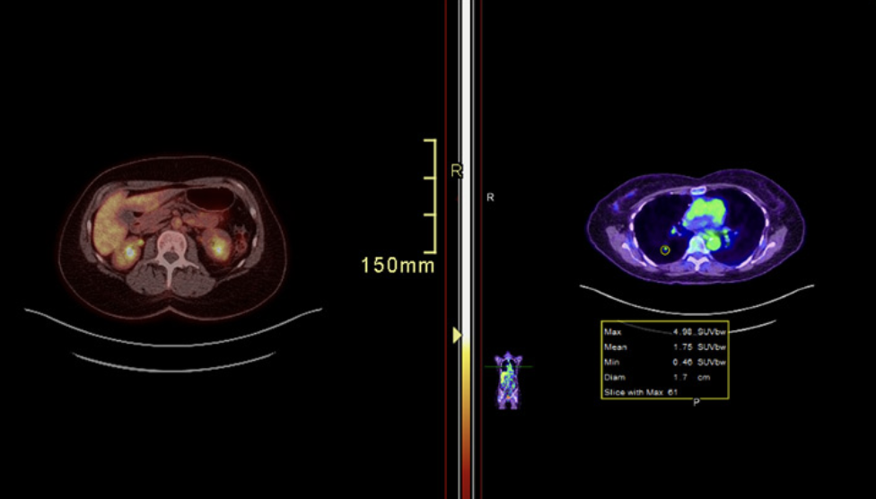
10123-pet-scan. (n.d.). Cleveland Clinic. https://my.clevelandclinic.org/health/diagnostics/10123-pet-scan
How it works
A radiotracer is built from a carrier molecule, such as a sugar, attached to a radioactive label like Technetium-99m or Fluorine-18. The carrier guides the label to targeted tissue, and radioactivity accumulates wherever biological activity is highest. As the tracer moves through the bloodstream, it's absorbed differently by healthy versus diseased cells — cancer cells or other highly active tissue tend to absorb more, creating "hot spots" the scanner can detect. Whatever isn't absorbed is naturally flushed out through urine. A PET scan is often combined with a CT scan, known as PET/CT, pairing PET's activity data with CT's detailed anatomy.
Radiotracer dose
For a standard adult scan using the most common tracer, FDG, doctors typically inject 10 to 20 millicuries (about 370 to 740 megabecquerels). The exact dose is calculated by nuclear medicine technologists based on body weight, balancing image quality against minimizing radiation exposure. The tracer itself contributes about 4 to 8 millisieverts of radiation; combined with the CT portion of a PET/CT scan, the total effective dose is usually 10 to 25 millisieverts — comparable to roughly 2 to 3 years of natural background radiation. A prepared radiotracer typically has a shelf life of about 8 hours.
Common types of PET tracers
| Tracer | Used for |
|---|---|
| FDG (fluorodeoxyglucose) | The most common PET tracer; shows how cells use sugar, which helps find or stage cancers that tend to consume more sugar |
| Gallium (e.g. gallium-68, DOTATATE) | Used to evaluate certain tumors or infections |
| Rubidium | Commonly used to assess blood flow to the heart |
| Other specialized tracers | Amyloid tracers for brain conditions like Alzheimer's disease; fluciclovine (Axumin) for prostate cancer |
When a PET scan is ordered
- Detecting cancer, including breast, lung, and thyroid cancer
- Evaluating coronary artery disease, heart attack, or other heart problems
- Assessing brain disorders such as brain tumors, epilepsy, dementia, and Alzheimer's disease
- Determining whether cancer has spread, whether treatment is working, or whether cancer has returned
- Evaluating damage after a heart attack or stroke and identifying areas of the heart needing treatment
During active cancer treatment, scans are typically performed after every 2 to 3 treatment cycles — roughly every 6 to 12 weeks — to assess response. After treatment ends, surveillance scans are usually done every 3 to 6 months at first, gradually spacing out to once or twice a year as recurrence risk drops. Medicare typically covers up to three follow-up PET scans per tumor type after initial treatment, though more can be covered with documentation of medical necessity. Scans are generally not scheduled more than once on the same day.
Preparing for a PET scan
- Share a current list of medications, vitamins, supplements, and any allergies with your provider.
- Let your provider know if you're pregnant, think you might be, or are breastfeeding.
- Avoid eating for six hours before the test — patients with diabetes should check with their provider, since this guidance may differ, along with any changes needed to diabetes medication or insulin use.
- Drink only water beforehand.
- Avoid caffeine for 24 hours before the test if being evaluated for a heart problem.
- Wear comfortable clothing and leave metal items — jewelry, eyeglasses, dentures — at home.
- Mention to your provider if enclosed spaces make you anxious; a mild sedative may be available to help you relax.
PET Scan Procedure
You'll receive an IV injection of a radiotracer that contains a safe amount of a radioactive drug. Here's what to expect:
- You'll sit in a chair for about an hour while the radiotracer moves through your bloodstream and gets absorbed by your organs and tissues. You won't be able to feel the radiotracer.
- If you're getting a PET/CT scan, you may also get an IV injection of a contrast dye. This dye helps irregularities stand out.
- You'll lie on an exam table that slides in and out of the PET/CT scanner. This scanner is shaped like a doughnut.
- You must remain still during the scan. Movement can blur the images.
- You'll hear buzzing and clicking sounds as the scanner takes images.
- A technologist will review the scans before you leave to ensure the images are in focus.
How long does a PET scan take?
The entire PET scan process takes about two hours. It can take up to 60 minutes for your body to absorb the injected radiotracer, during which you'll need to sit quietly and limit your movements. The actual PET scan takes about 30 minutes. After the test, you'll need to wait while the technologist reviews the scans to ensure the images are clear.
Risks and side effects
PET scans are generally safe and rarely cause problems. The radiation dose is low and doesn't stay in the body long — drinking plenty of water afterward helps flush it out faster. Risk is mainly a concern in a few specific situations: pregnancy or breastfeeding, since radiation could affect a fetus or pass through breast milk; a rare allergic reaction to the tracer or to CT contrast dye; and diabetes, which can affect how the body absorbs the sugar-based tracer and may require diet or medication adjustments beforehand.
After the scan
Most people resume normal activities right away. The tracer clears the body within a few hours, and drinking fluids helps speed that up. Mild discomfort at the injection site is possible but uncommon. A radiologist trained in PET imaging reviews the results and sends a report to the ordering provider, typically within 24 to 48 hours.
Understanding the results
- Normal — no signs of disease or irregularities.
- Abnormal — findings suggesting infection, inflammation, or cancer.
Results can also show how a disease is progressing or how well a treatment is working. Contact your healthcare provider if you notice dizziness, nausea, difficulty breathing, or worsening pain at the injection site after the scan.