fNIRS Overview
Functional near-infrared spectroscopy (fNIRS) is an emerging brain imaging technology, less mainstream than techniques like MRI, CT, PET, or ultrasound. It's a portable, non-invasive method that uses low levels of non-ionizing light to record changes in cerebral blood flow through optical sensors placed on the surface of the scalp.
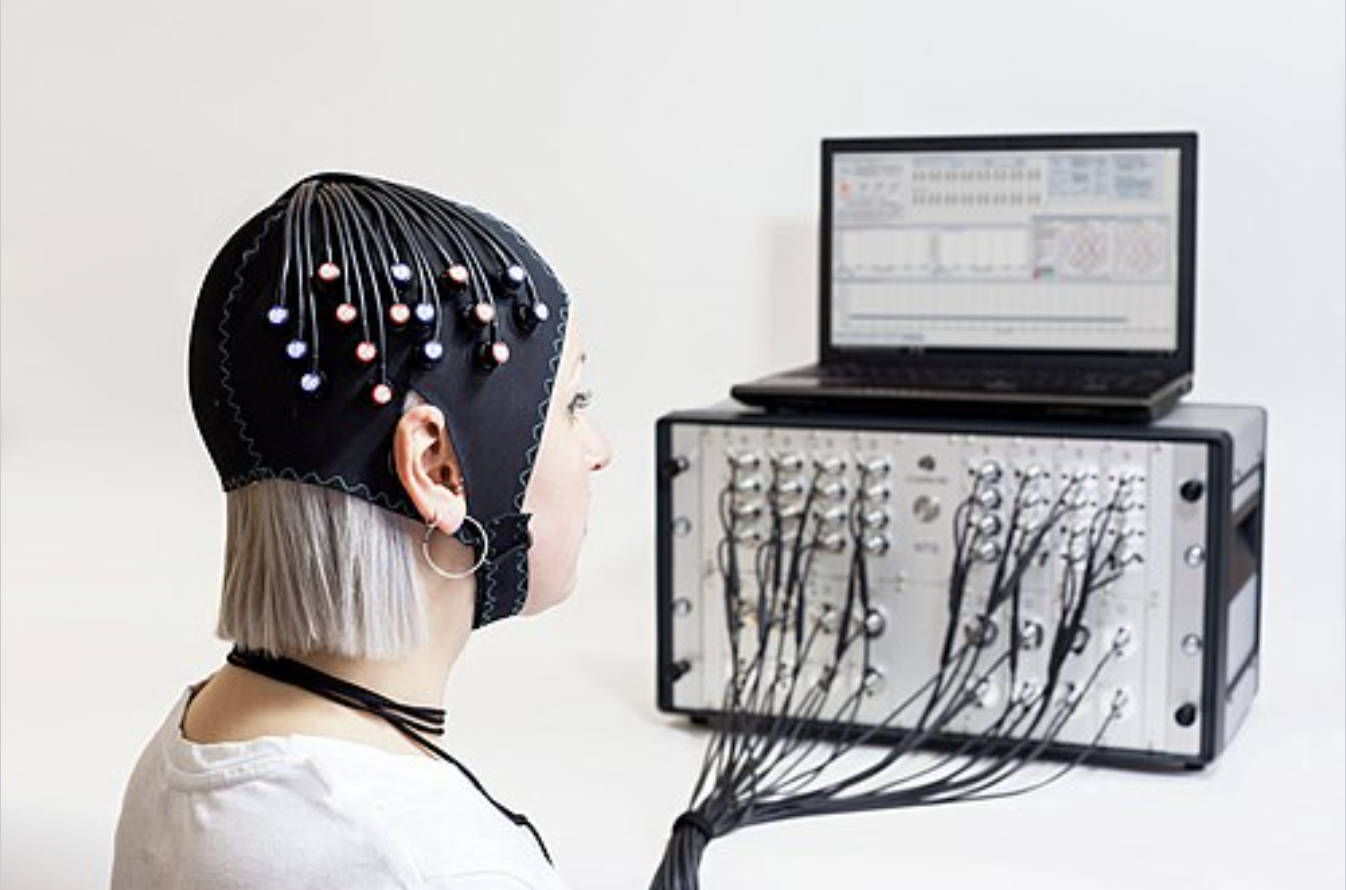
fNIRS with a Gowerlabs NTS system. (2019, November). Wikipedia. https://en.wikipedia.org/wiki/Functional_near-infrared_spectroscopy#/media/File:Blonde_fNIRS_lady.jpg

How it compares to other brain imaging
Like fMRI, fNIRS measures changes in blood oxygenation as a way of inferring brain activity, but it does so without magnetic fields or ionizing radiation. Compared with fMRI or PET, fNIRS is relatively inexpensive, small, and portable — signals travel through flexible fiber optic cables from a head cap worn by the participant to the recording instrument. That portability makes it possible to record brain activity during real-world tasks, such as walking, standing, or navigating balance perturbations, situations that are difficult or impossible to study inside an MRI or PET scanner. fNIRS is limited to cortical brain activity, since near-infrared light can only penetrate the outer layers of the brain, and it's sometimes combined with EEG for a fuller picture of brain activity.
Preparing for an fNIRS session
- Head measurement — participants are typically asked to measure their (or their child's) head with a measuring tape beforehand, so the correct cap size can be selected — not too tight, not too loose.
- Clothing — fNIRS has no clothing restrictions like MRI does, though hair is usually best left down, without braids or ponytails, so the cap can make good contact with the scalp. If an MRI is scheduled the same day, clothing without metal (no jewelry, buckles, or hair accessories) is recommended. Glasses are fine for fNIRS but not for MRI, so participants with an MRI scan the same day may need MRI-safe glasses or contacts instead.
fNIRS Scan Procedure
Before your appointment
Measure your head: before your scheduled NIRS session, you will be asked to measure your or your child's head using measuring tape. We need this measurement to choose the correct NIRS cap size so that it is not too tight or too loose. We need this information at least one day before your appointment, so email us the head size as soon as you can. Please watch our instructional video on how to properly measure your head.
Dress comfortably: NIRS does not have clothing restrictions like MRI scans, so feel free to wear any kind of outfit you like. If your study does have specific clothing guidelines (e.g. exercise studies), the research coordinator will let you know ahead of time. For those with long hair, leave your hair down without braids or ponytails if possible. If you will have an MRI scan on the same day, wear clothes that are comfortable and do NOT contain any metal (no jewelry, hair accessories, buckles, or belts). Glasses are fine to use with NIRS, but you will not be able to use them in the MRI scanner. Please let your research coordinator know your prescription ahead of time so they can prepare MRI-safe glasses or use contact lenses instead.
During the scan
Cap set-up and signal optimization: before the NIRS scan, we will need to place the cap on your head. The cap contains light sources and detectors that are placed around the head. It is not painful, and we can adjust the cap to alleviate any discomfort. Putting on the cap only takes a minute. Once the cap is on, we will need to run a signal optimization to make sure the sources and detectors have a strong signal. Since light needs to shine into the head from the scalp, we may need to adjust the optodes by using a q-tip to push hair or wires out of the way. In cases when hair is very thick or dark, we may use gel. The gel is water-based, so it is not sticky and dries out quickly, so you won't even know it was there at the end of the session.
NIRS scan: once we have a good signal, it's time to start the NIRS scan. Each study has different tasks for their scan. Common examples include playing games on the computer or having a conversation with someone else. We even have a project where participants ride a bike outside while getting a scan! Your study's research coordinator should have already provided you with details on what to expect during the NIRS scan. Most scans last about 20-40 minutes, but may be shorter or longer depending on the project.
After your appointment
After your session, a researcher will take you or your child to their next study agenda item. This could be an MRI scan, answering questionnaires, or other study-specific activities. Or NIRS may be the last thing you need to do for the study appointment, and you will be given information about compensation and next steps. We greatly appreciate your participation!
What fNIRS can reveal
By tracking relative changes in oxygenated and deoxygenated hemoglobin, fNIRS lets researchers infer metabolic activity and, by extension, brain activation, since cerebral blood flow increases in the areas of the brain being used for a given task — a relationship known as neurovascular coupling. This has made fNIRS especially useful for studying ambulatory motor learning, balance, and vestibular function — questions that are hard to study with imaging methods that require the participant to stay still inside a scanner.