Ultrasound Imaging
An ultrasound is an imaging test that uses high-frequency sound waves to create real-time pictures or video of soft tissues inside the body. It is a noninvasive procedure, also called sonography or ultrasonography, and the resulting image is known as a sonogram. Exams are performed by a doctor or a trained specialist called an ultrasound technologist or sonographer.
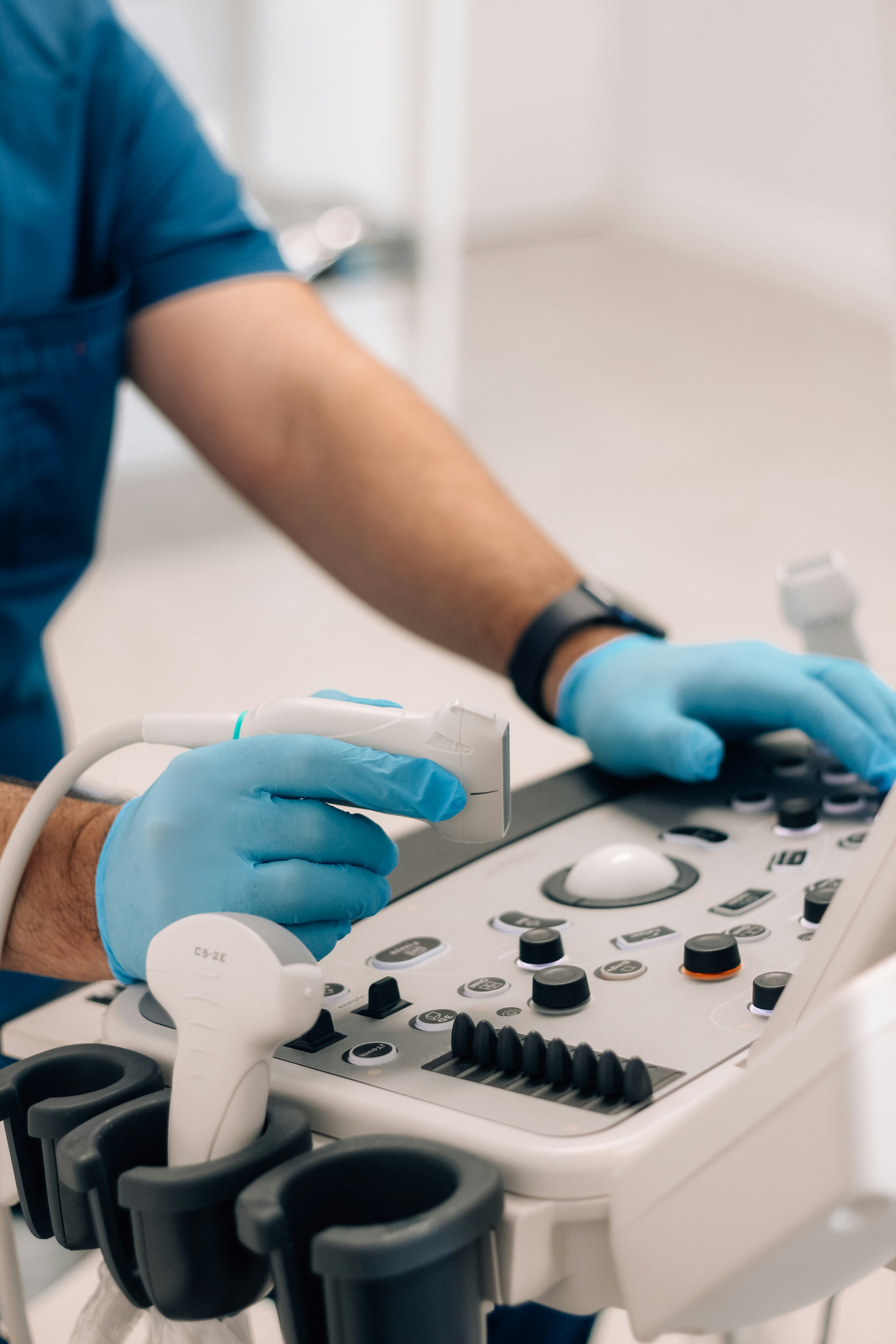
Sher, E. (2023, August). A person in blue gloves operating a machine. Unsplash. https://unsplash.com/photos/a-person-in-blue-gloves-operating-a-machine-ZL-xUYvkaKM
Main categories of ultrasound
- Pregnancy ultrasound — confirms pregnancy, estimates due date, and checks fetal growth.
- Diagnostic ultrasound — views internal organs and tissues to check for problems, such as abdominal or breast ultrasound.
- Ultrasound guidance for procedures — helps guide needles, catheters, or other instruments during a procedure, such as a biopsy or nerve block.
2D, 3D, and 4D imaging
Most ultrasound images are two-dimensional, producing a flat picture of internal structures. Three-dimensional ultrasound adds depth and angle information that a 2D image can't capture, and four-dimensional ultrasound is essentially 3D imaging shown live, in motion.
How it works
A handheld device called a transducer, or probe, is passed over the skin or inserted into a body opening. A thin layer of gel is applied first so the sound waves transmit cleanly from the probe into the body. The probe converts an electrical current into high-frequency sound waves, which are inaudible to the patient. These waves bounce off internal structures and return to the probe, which converts them back into electrical signals. A computer then turns that signal pattern into the real-time image or video seen on screen.
Ultrasound Procedure
Ultrasounds that involve applying the transducer over your skin (not inside your body) follow these general steps:
- You'll lie on your side or back on a comfortable table.
- The ultrasound technologist will apply a small amount of water-soluble gel onto your skin over the area to be examined. This gel doesn't harm your skin or stain your clothes.
- The technologist will glide a handheld transducer or probe over the gel to get images inside your body.
- The technologist may ask you to be very still or to hold your breath for a few seconds to create clearer pictures.
- Once the technologist has gotten enough images, they'll wipe off any remaining gel from your skin, and you'll be done.
- If the transducer is going inside your vagina or rectum, it follows the same general steps above, but with one exception. Before the exam, your provider will put a sterile, lubricated cover on the transducer, so the transducer goes in more easily.
For internal exams, a sterile, lubricated cover is placed over the transducer beforehand. Skin-surface ultrasounds are generally painless, while internal exams may be uncomfortable but shouldn't hurt. A full exam usually takes 30 minutes to an hour.
Common uses
| Procedure | Purpose |
|---|---|
| Abdominal ultrasound | Visualize abdominal tissues and organs |
| Bone sonometry | Assess bone fragility |
| Breast ultrasound | Visualize breast tissue |
| Doppler fetal heart rate monitor | Listen to the fetal heartbeat |
| Doppler ultrasound | Visualize blood flow through vessels, organs, or other structures |
| Echocardiogram | View the heart |
| Fetal ultrasound | View the fetus during pregnancy |
| Ultrasound-guided biopsy | Collect a tissue sample |
| Ophthalmic ultrasound | Visualize ocular structures |
| Ultrasound-guided needle placement | Guide needles into blood vessels or other target tissue |
Imaging modes
- A-mode (Amplitude) — the earliest mode, showing returning echoes as a one-dimensional graph, useful for measuring tissue thickness or distance. Largely superseded by other modes today.
- B-mode (Brightness) — represents echo amplitude as dots or pixels of varying brightness, forming the standard real-time image seen on most systems.
- M-mode (Motion) — displays echo data for a single image line over time, commonly used in echocardiography to track valve and chamber motion through the cardiac cycle.
Doppler modes
Doppler ultrasound uses shifts in sound wave frequency caused by moving structures (typically blood) to measure direction and velocity of flow.
| Mode | How it works | Trade-off |
|---|---|---|
| Pulsed Wave (PW) | Alternates emitting a pulse and listening for its echo at a specific depth | Depth-specific, but limited in the flow velocities it can accurately measure |
| Continuous Wave (CW) | Emits and receives sound continuously along the beam path | Measures higher velocities accurately, but can't isolate a specific depth |
Doppler data can then be displayed as spectral Doppler (a graph of velocity over time), colour Doppler (direction and relative velocity shown as colored pixels over the B-mode image), or power Doppler (a more sensitive, single-color display of flow strength without directional information).
Safety
Ultrasound has an excellent safety record and, unlike X-rays or CT scans, doesn't use ionizing radiation. It carries no known risk of birth defects or developmental disorders. Ultrasound energy can, however, slightly heat tissue and in some cases produce small gas pockets in body fluids (cavitation); the long-term significance of these effects is still unknown. Because of this, professional guidance encourages prudent use during pregnancy, and discourages ultrasound performed solely for non-medical purposes, such as keepsake fetal videos, unless it occurs during an already medically indicated exam. All ultrasounds should be performed by a trained professional.
Getting results
Turnaround time varies by exam type. Some results are available quickly, while others take several days for a radiologist to review. Once reviewed, a report is sent to the provider who ordered the exam, and that provider shares the findings with the patient.